
Maths plus medicine equals new imaging innovation
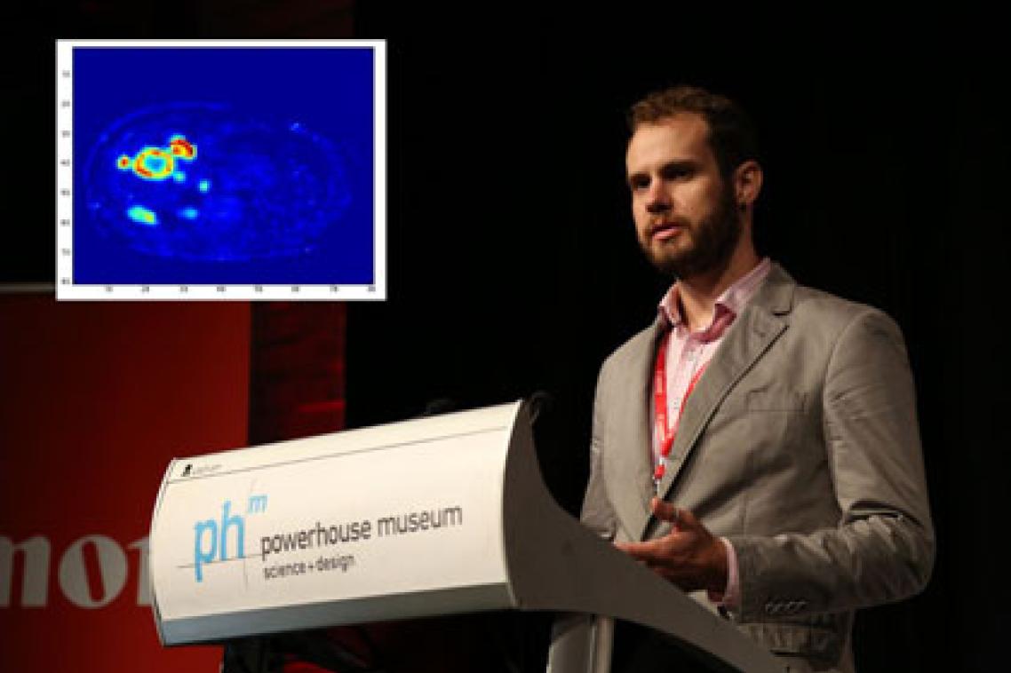
A medical student from The University of Queensland has applied his background in mathematics to the field of nuclear medicine to improve liver imaging methods, which may improve diagnosis time and save patients undergoing unnecessary surgery.
Third year medical undergraduate Charles Baker has developed a mathematical model that can be programmed into existing scanning equipment to enhance images of patients’ livers.
Mr Baker said the model uses variables specifically relating to the structure of the liver to help nuclear medicine specialists better differentiate between healthy and damaged liver tissue.
“This means we can improve the output of existing scanning equipment to provide better image quality for nuclear medicine specialists,” Mr Baker said.
“The resulting images demonstrate higher contrast between healthy liver tissue and unhealthy liver tissue, such as malignant tumours.”
The mathematical model is undergoing clinical appraisal in the Nuclear Medicine Department at the Royal Brisbane and Women’s Hospital (RBWH) and Mr Baker said initial feedback had been encouraging.
“We hope that practical testing of the model across a larger number of images will help identify how it will improve clinical decision-making and patient outcomes.”
“For example, scans using the mathematical model may help to more clearly identify areas of dead tumour tissue and active tumour tissue. Clinicians can use this information to better target treatments.”
The improved contrast would also help specialists to more easily interpret scans, saving time.
“Most importantly, the higher contrast scans might save people from undergoing unnecessary surgical procedures,” Mr Baker said.
He is now working on mathematical models to improve imaging in other parts of the body, such as the brain.
Mr Baker developed the model under the supervision of Dr Nicholas Dowson and Professor Steven Rose, from The Australian eHealth Research Centre, CSIRO, and Dr Paul Thomas, Associate Director of Specialised PET Services, Department of Nuclear Medicine, RBWH.
Mr Baker was recently recognised for his work with the Undergraduate Prize in Canon Australia’s Extreme Imaging Competition.
Media contact: Faculty of Medicine and Biomedical Sciences, Kate Gadenne, 0438 727 895, k.gadenne@uq.edu.au.
Topics
Related articles

Feed the children: food provision in early childcare falls short
