Zooming in: UQ photo contest captures science at the nanoscale
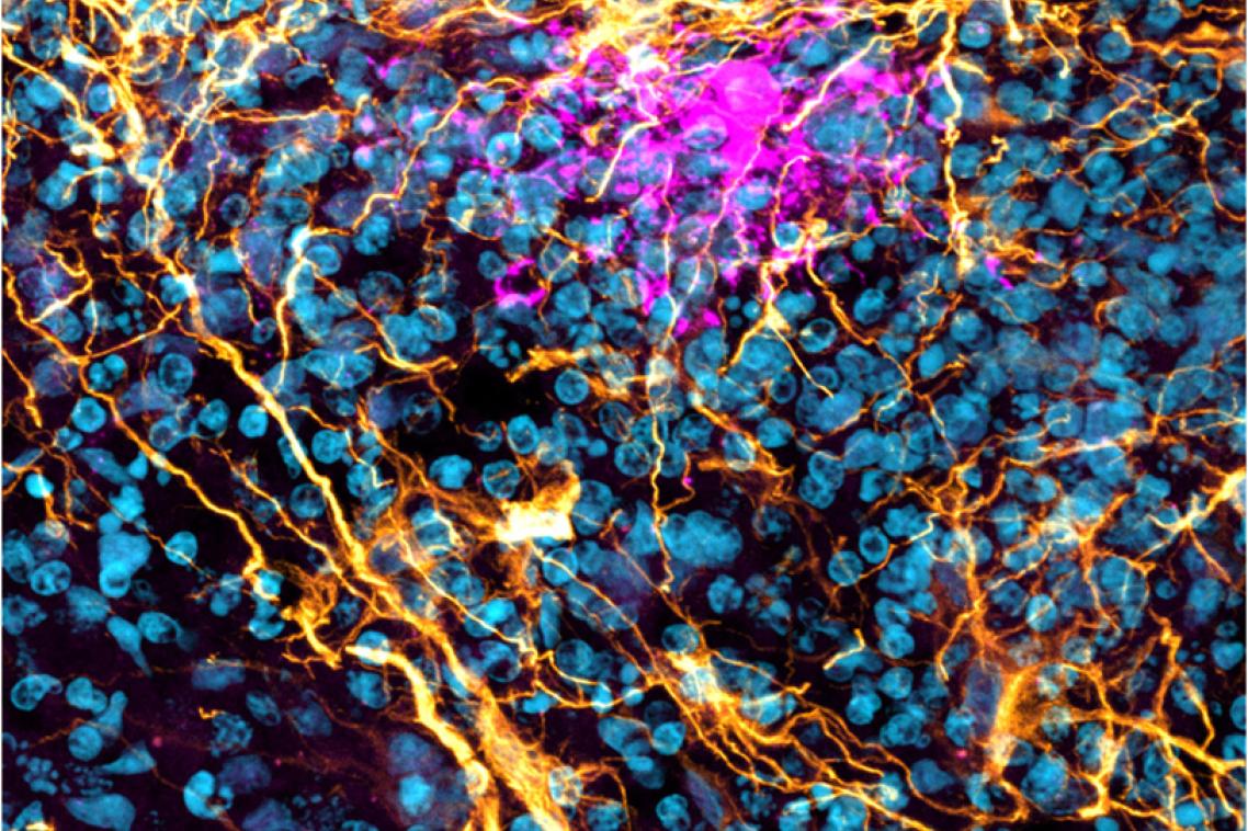
The winning image in this year's AIBN photo competition for National Science Week - an osteocyte suspended in a 3D biomimetic hydrogel system, captured by Shiva Muthuswamy.
(Photo credit: Shiva Muthuswamy / The University of Queensland. )
To explore the nanoscale is to peer into another world, a place where the rules of science bend and the unseen comes alive.
Luckily, zooming in is what researchers at UQ’s Australian Institute for Bioengineering and Nanotechnology (AIBN) do best. And they love to show off the things they find.
Each year during National Science Week AIBN researchers compete to see who can take the best photo using the powerful imaging technology found throughout the institute.
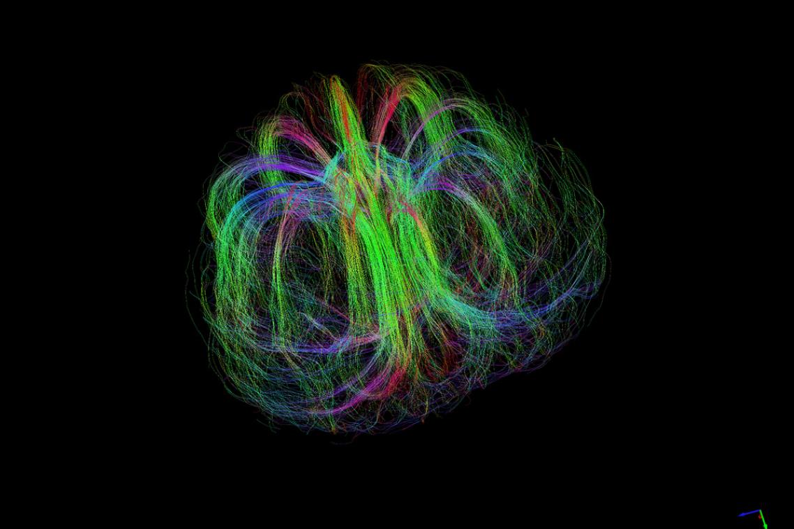
Researchers at the AIBN have access to some incredible imaging technology. Here, diffusion MRI tractography has been to map a developing Burderkin plum, specifically the fibrous structures that supply various nutrients.
(Photo credit: Nyoman Kurniawan / The University of Queensland )
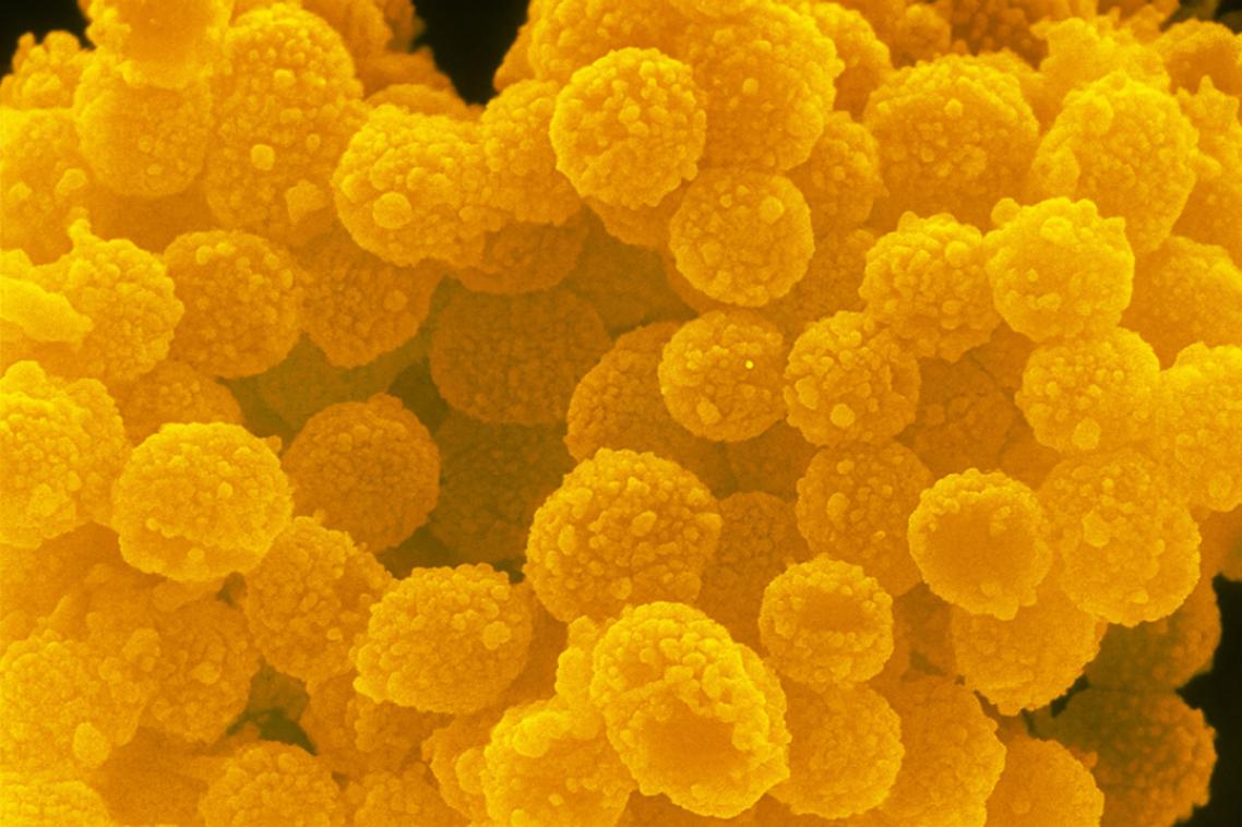
Sea anemone or metal organic framework? Hint: probably best this doesn’t go in the ocean.
(Photo credit: Silvia Chowdhury / The University of Queensland )
Don’t let this caramel popcorn fool you this striking image captured using a (SEM) actually showcases SiO₂@Au/Au nanostructure. What looks delicious is, in fact, the intricate beauty of nanoscale engineering, where science meets art.
(Photo credit: Junaid Munawar / The University of Queensland )
Brain organoids are tiny, synthetic representations of real human brains. This section of a human brain organoid shows the star-shaped glial cells known as astrocytes, aged 140 days.
(Photo credit: Bahaa Al-mhanawi / The University of Queensland )
Not a palm leaf or the inside of a bottle, but a fluorescein isothiocyanate lipids vacuum-dried down to form a multilayer and imaged on a 10x fluorescence microscope.
(Photo credit: Aidan Thiele / The University of Queensland )
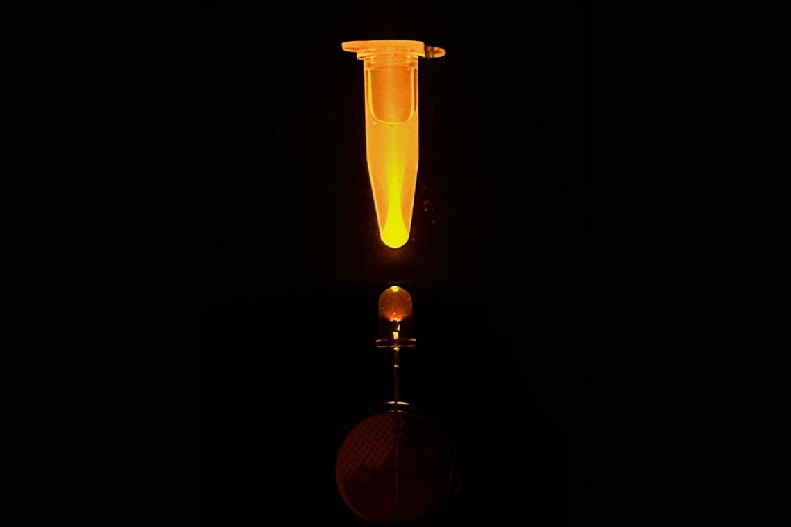
Extracellular matrix mediated biophysical cues dynamically modulate the behaviour and functions of osteocytes. A lensing effect is produced as LED light passes through the base of a clear sample tube, revealing fluorescently stained proteins in solution.
(Photo credit: Will Anderson / The University of Queensland )
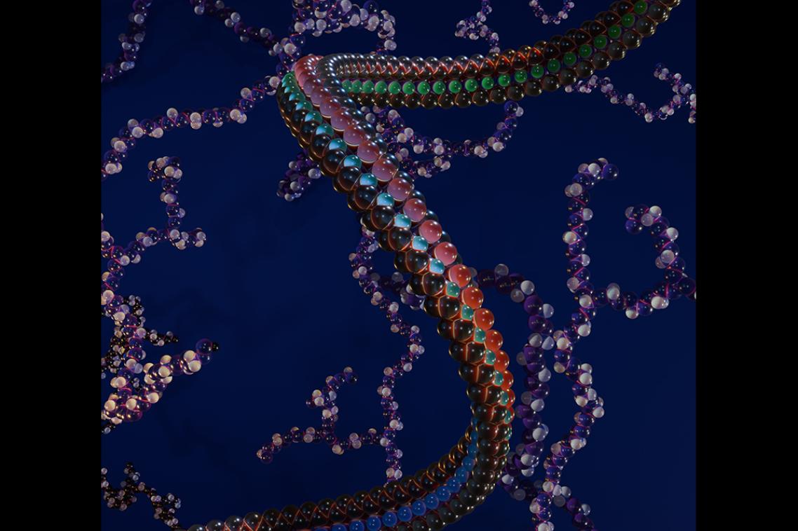
A showcase of digital PolyNIPAM and polyethyleneimine models, created from real polymer simulations via the PolyConstruct platform.
(Photo credit: Ada Quinn / The University of Queensland )
Once again, this year’s winners have shown that some of the most impressive sights in science often start out as specks on a glass plate.
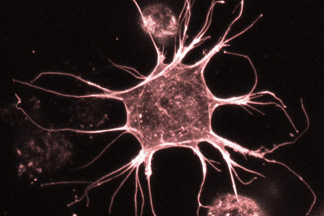
The judges’ pick for the 2025 AIBN Image Contest was taken by Shiva Muthuswamy – an osteocyte suspended in a 3D biomimetic hydrogel system.
Shiva said ten to fifteen minutes under a confocal microscope was all he needed to capture this most abundant type of bone cell.
“My research aim is to deepen our understanding of osteocyte’s mechanobiology,” Shiva said.
“If we can learn more about these cells, we can learn more about the decline in bone density we experience as we age, and how we might prevent that.”
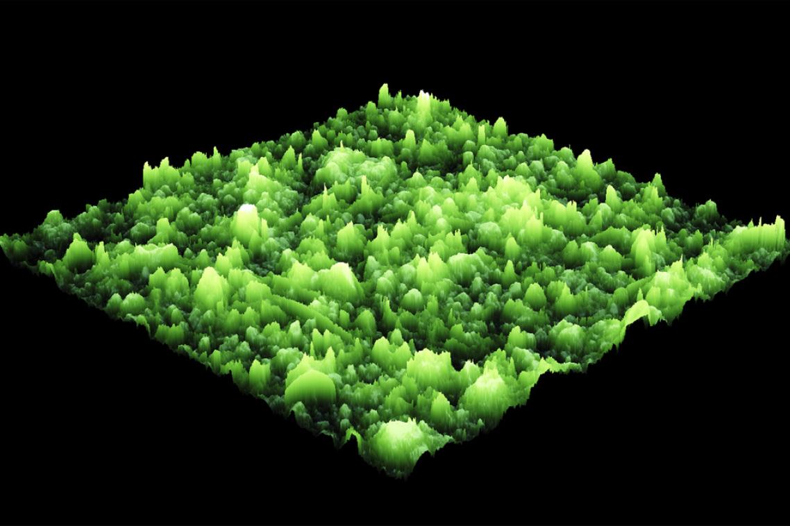
Zinc electrode after cycling in an aqueous battery.
(Photo credit: Yiqing Wang / The University of Queensland )

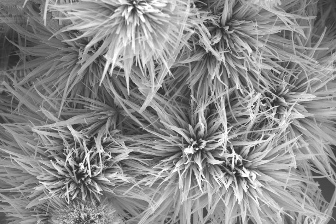
Here’s one for the family Christmas card. Fern-like copper nanocatalyst prepared by electrodeposition.
(Photo credit: Yusuf Valentino Kaneti / The University of Queensland )
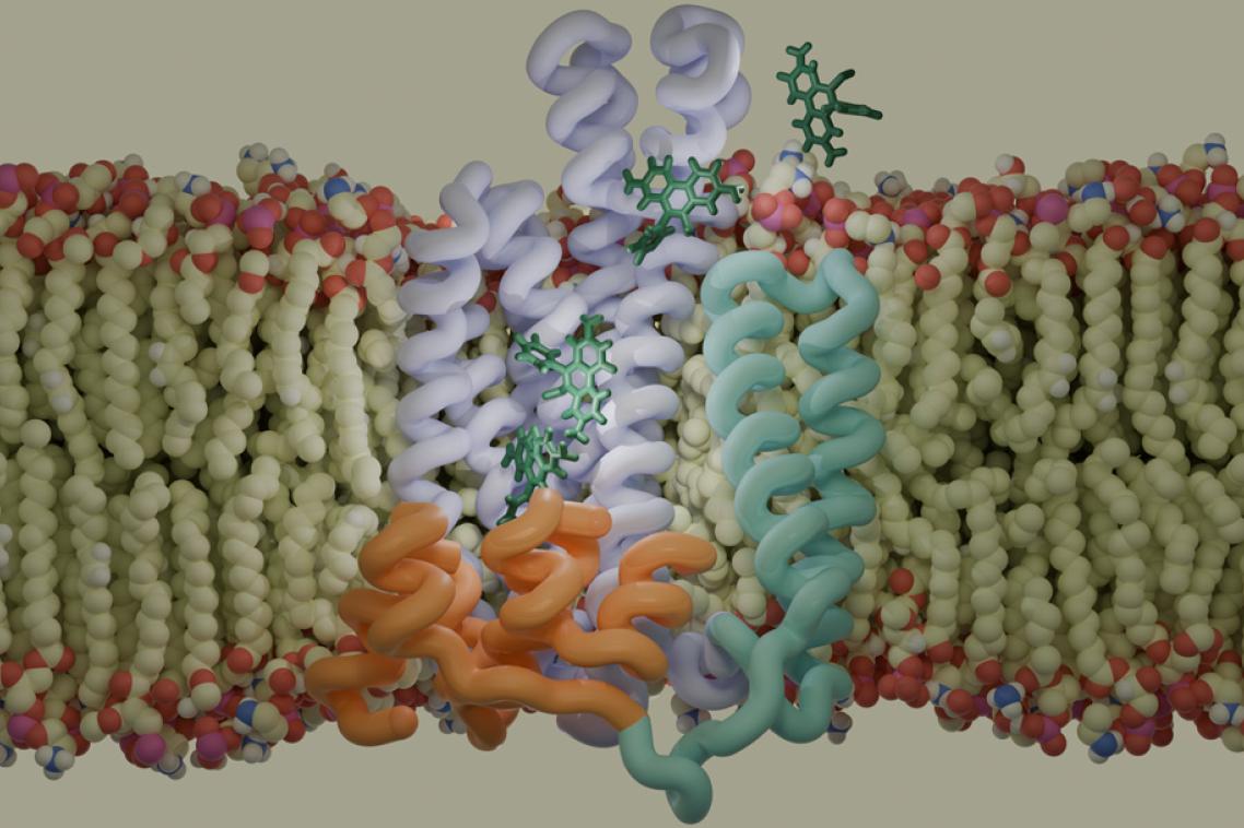
It’s not all Petrie dishes and scanning tech at the AIBN. Here a molecular dynamics simulator has been used to represent a QacA multidrug efflux pump transporting Ethidium bromide across the cell membrane in Methicillin-resistant Staphylococcus aureus.
(Photo credit: Patrick Sutton / The University of Queensland )
Most people would probably never guess what these are in a million years. (It’s a 3D scan of finger limes).
(Photo credit: Image by Nicole Atcheson / The University of Queensland )

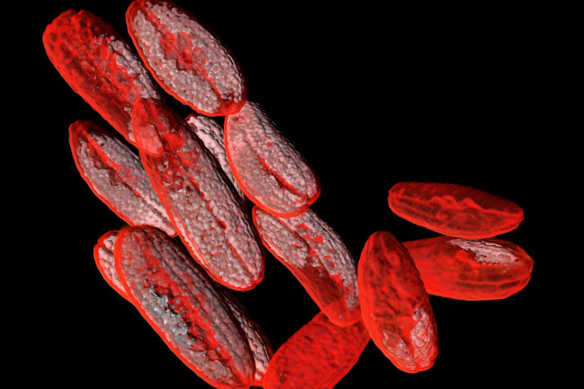
AIBN researchers aren’t typically bodybuilders, but nobody beats them when it comes to growing muscles. Pictured here is myotube grown in a dish using muscle stem cells.
(Photo credit: Melinder Gill / The University of Queensland )
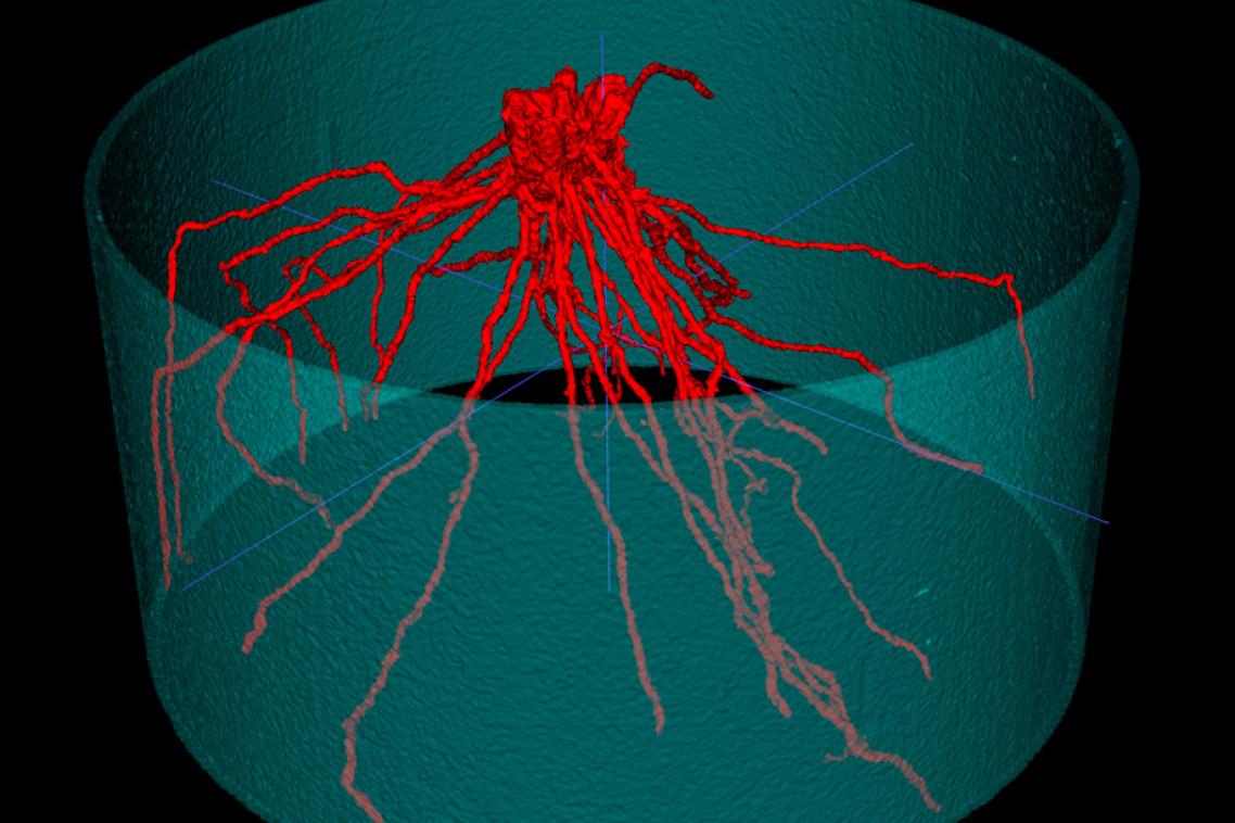
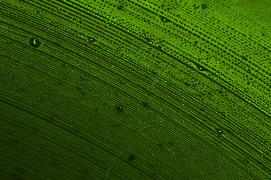
A microCT scanner uses powerful 3D imaging techniques to see inside objects, slice by slice. This shot represents the growth variation in a wheat root in different soil strengths.
(Photo credit: Gary Cowin/The University of Queensland )
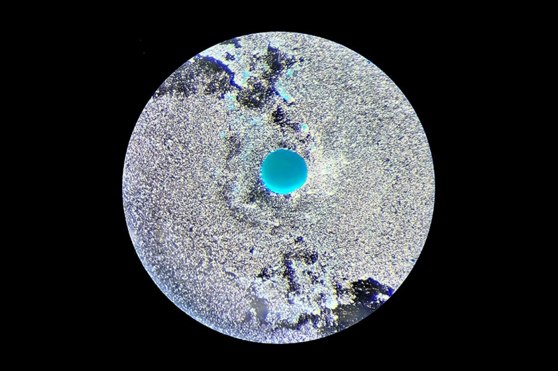
Like distant exoplanet of an unknown world, this image unveils the incredible regenerative potential of dental pulp stem cells. The vibrant blue sphere specifically illuminates the cartilage-like matrix these cells have formed, confirming successful chondrogenesis.
(Photo credit: Desi Veleva / The University of Queensland )
In second place for this year’s image competition was Dr Nyoman Kurniawan, who used diffusion MRI tractography to demonstrate the fibrous structures that supply nutrients throughout a Burdekin plum.
Finally, taking home the people’s choice prize in 2025 was Silvia Chowdhury, whose use of the scanning electron microscope on this metal organic framework attracted the most votes from friends and colleagues.
This year’s image competition was once again featured by Guardian Australia.
Related articles
New ultrasound imaging to map drug delivery into the brain